TenEx Core
TenEx Core
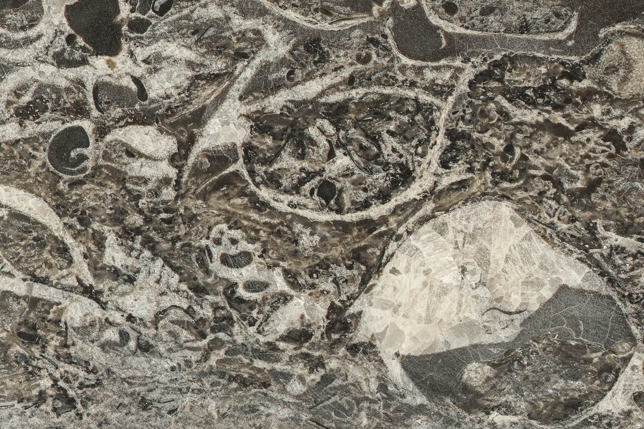
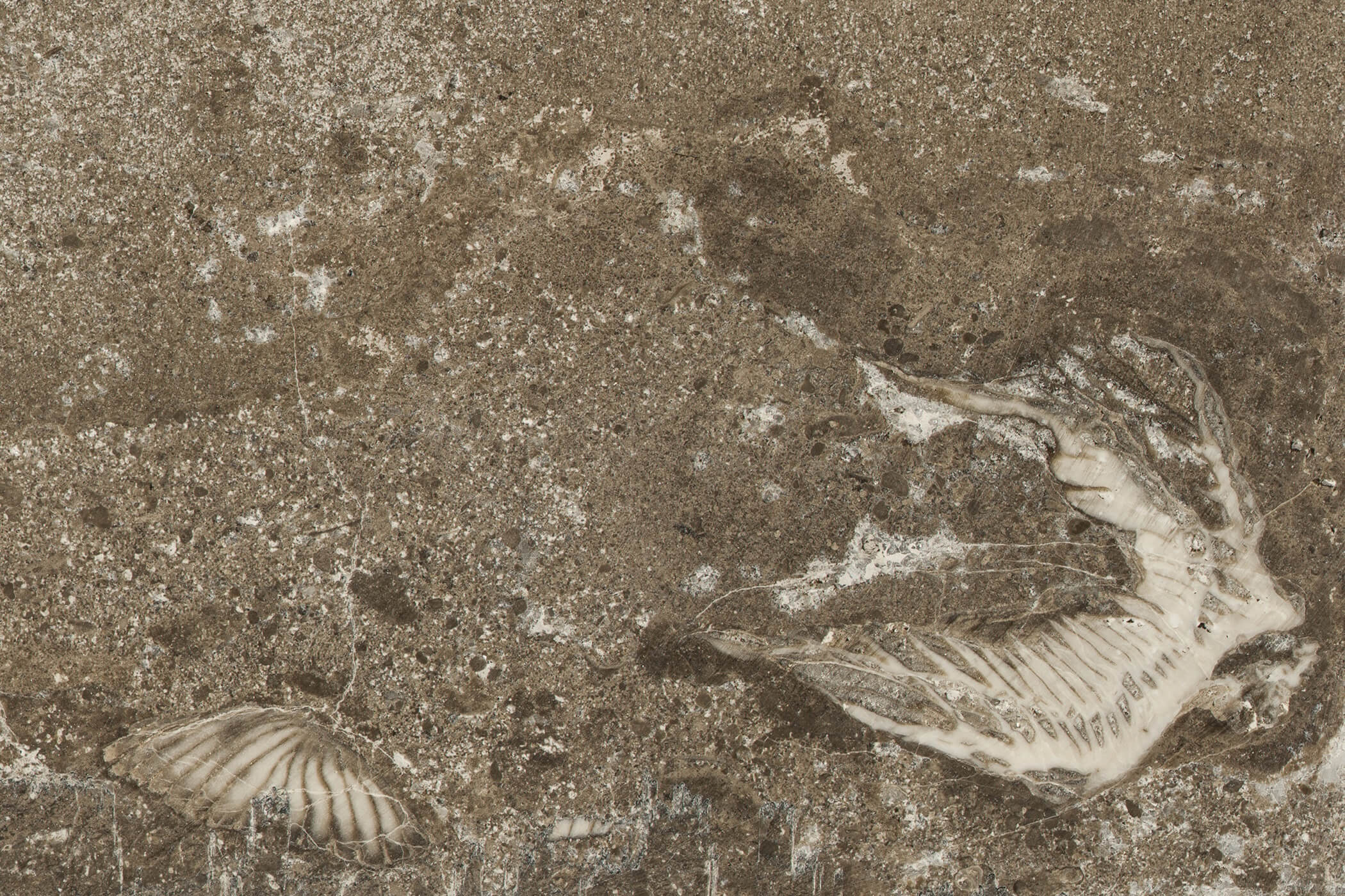
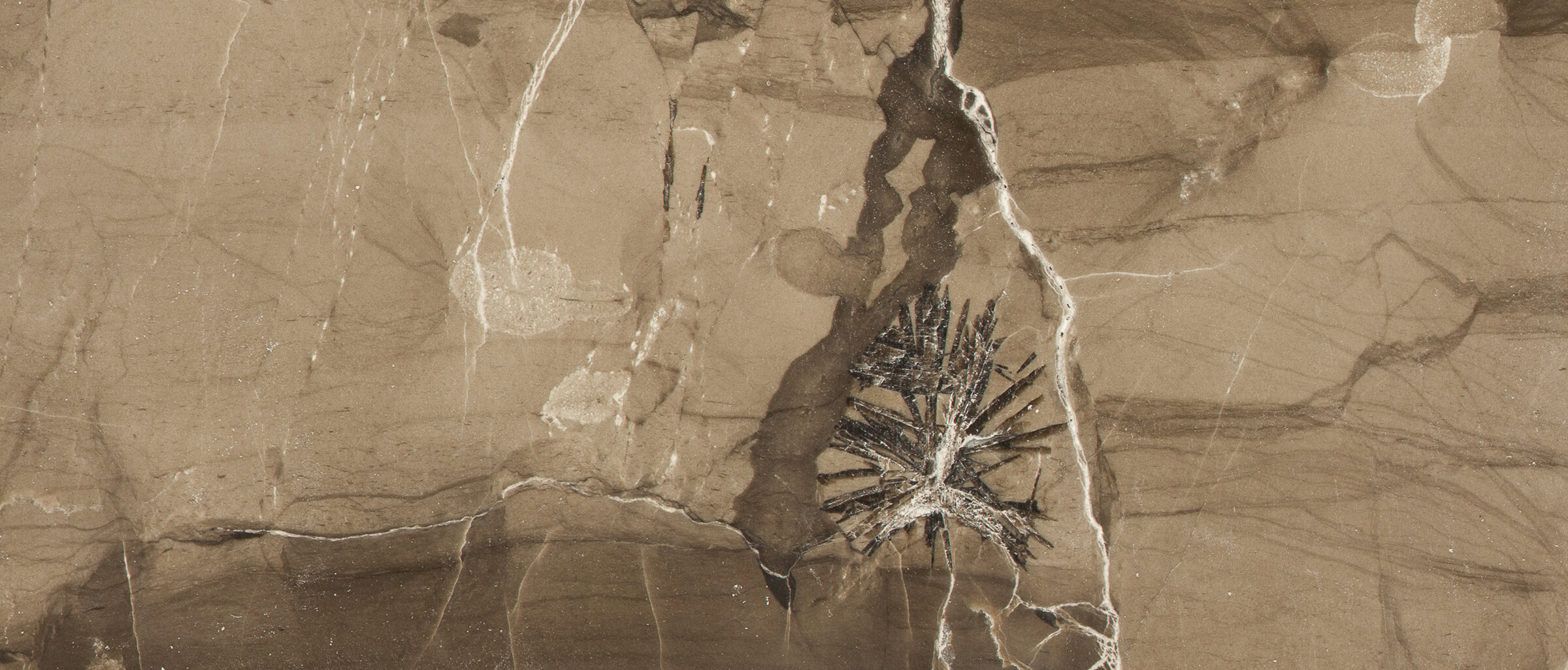
PetroArc imagery is designed to provide a permanent archive of your core in pristine condition. Now, all your core access is at your fingertips, resulting in cost reduction and increased production. CORSystem can facilitate the use of existing legacy images as well.

Standard core imagery in the oil and gas industry is 300dpi resolution. TenEx standard imagery begins at 1,360dpi up to a maximum of 11,000dpi. This imagery can be coupled with our software suites to perform well characterization.

MicroPics
MicroPics
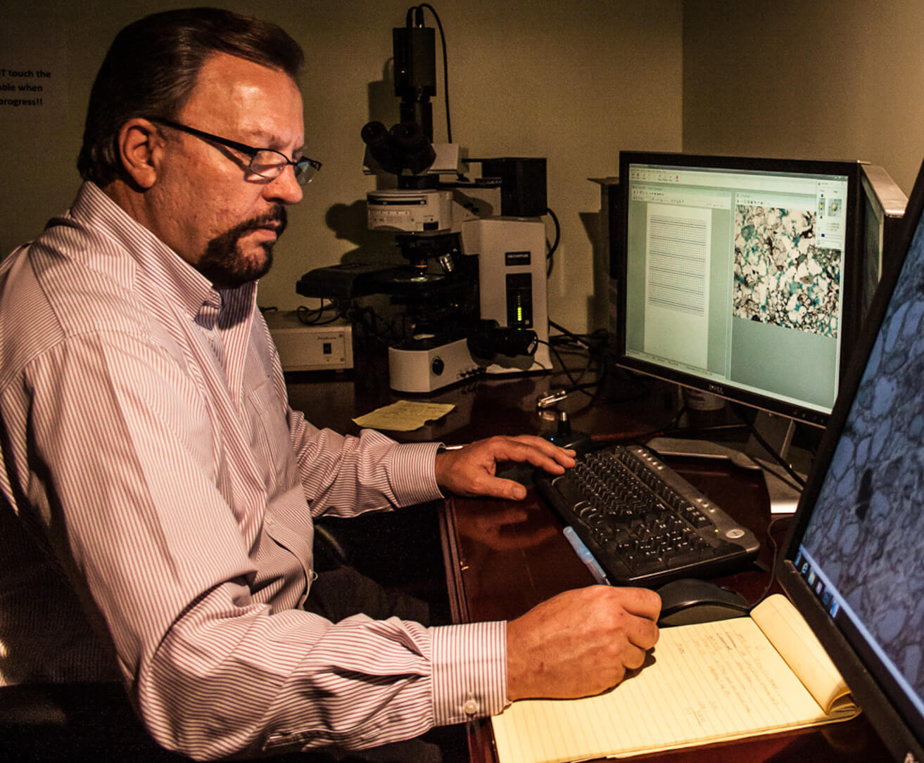
MicroPics are digital petrographic thin section images. Thin Sections play an integral part in most well characterization projects. Combining MicroPics with MicroPet software will allow users to have the most efficient access to their thin sections from anywhere. MicroPics are used in conjunction with our MicroPet software to perform grain size, sorting, and grain contacts, sedimentary structures (laminations and bioturbation), framework grain composition, authigenic minerals, and types and distribution of macro porosity.

Access your archived data quickly and efficiently without leaving your desk.

Immortalize your samples and data with our protocols that meet and surpass industry standards. Gain the advantage you need for your next project from anywhere in the world.
Contact Us
HD Plugs & Well Cuttings
High definition core/sidewall plugs imaged up to 80X magnification under white and/or UV light provide clear visibility of individual pores. Core plugs and sidewall plugs play an integral part in most well characterization projects. By combining HDPlugs with our software suite, well viability can be estimated with grain size analysis.

For every sidewall core taken in a field there may be hundreds of cuttings samples. Core cuttings can be integrated with mud-logger’s notes to provide an often untapped source of valuable information for reservoir characterization. Cuttings are routinely sampled, packed into envelopes, and stored somewhere in a box. If needed, they will be difficult, if not impossible to locate. Our archiving methodology provides instant, one-click access to any sample or group of samples.

Histology and Neuroanatomy
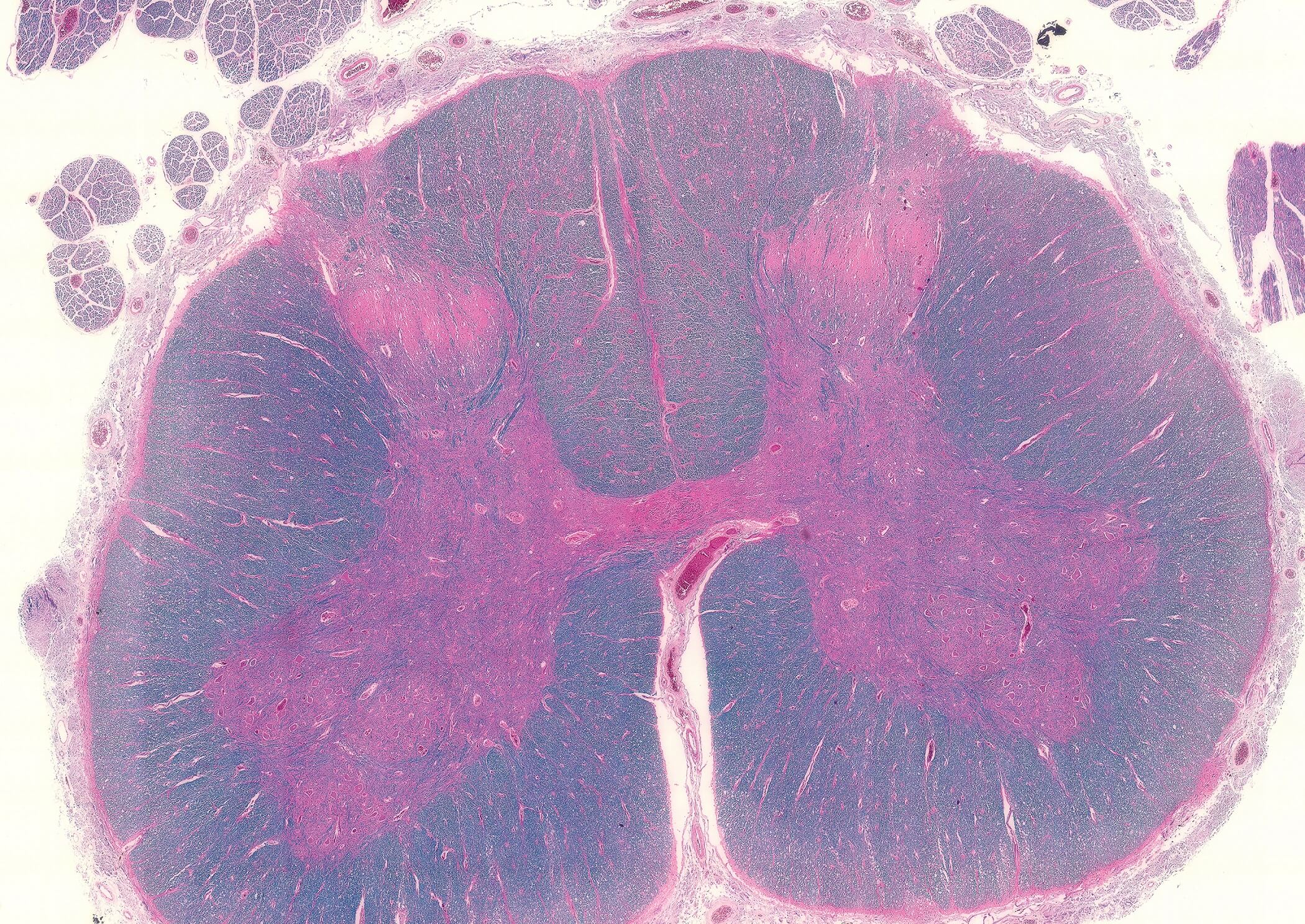
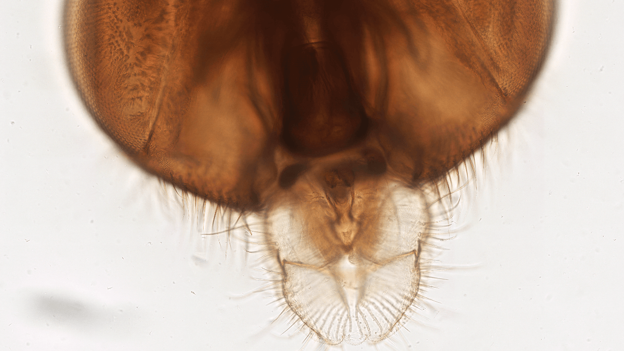
Visualize and investigate specimens using traditional light microscopy techniques. Identify bacteria, carry out cellular structure studies, or explore neuroanatomy. If you can send us your samples, we can image them.

Check out a small selection of virtual slides and virtual microscopy specimens we create for our clients in our gallery!

View Full Gallery