NERV-002 | Slides maybe purchase individually or as custom collections. If you wish to purchase 25 or more virtual slides, discounts will be automatically applied according to incremental package sets of 25, 50, 100, 200, or unlimited.
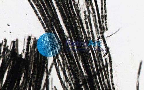
Nerve Fibers, 200X Osmium Tetroxide
Product Description
A nerve fiber is a threadlike extension of a nerve cell and consists of an axon and (in some cases) a myelin sheath. There are nerve fibers in the central nervous system and peripheral nervous system. In the central nervous system (CNS), myelin is produced by oligodendroglia cells. Schwann cells form myelin in the peripheral nervous system (PNS). Schwann cells can also make a thin covering for an axon which does not consist of myelin (in the PNS). A peripheral nerve fiber consists of an axon, myelin sheath, Schwann cells and its endoneurium. There are no endoneurium and Schwann cells in the central nervous system. OsO4 is a widely used staining agent used in transmission electron microscopy (TEM) to provide contrast to the image. As a lipid stain, it is also useful in scanning electron microscopy (SEM) as an alternative to sputter coating. It embeds a heavy metal directly into cell membranes, creating a high electron scattering rate without the need for coating the membrane with a layer of metal, which can obscure details of the cell membrane. In the staining of the plasma membrane, osmium(VIII) oxide binds phospholipid head regions, thus creating contrast with the neighbouring protoplasm (cytoplasm). Additionally, osmium(VIII) oxide is also used for fixing biological samples in conjunction with HgCl2. Its rapid killing abilities are used to quickly kill live specimens such as protozoa. OsO4 stabilizes many proteins by transforming them into gels without destroying structural features. Tissue proteins that are stabilized by OsO4 are not coagulated by alcohols during dehydration. Osmium(VIII) oxide is also used as a stain for lipids in optical microscopy. OsO4 also stains the human cornea.


